

Knee Dislocations
Case : A 26-year old male is brought to the emergency department (ED) after a motor vehicle collision. The vehicle sustained a frontal impact with a lamp post. In the trauma bay, he is conscious and alert. He is complaining of severe knee pain and reports having felt a sudden tearing and popping sensation in his knee during the impact.
Take Home Points:
- Knee dislocations are limb threatening injuries
- The physician should maintain a high level of suspicion as the presentation may be subtle
- Knee dislocation often happens with a high velocity mechanism but in patients with morbid obesity knee dislocation is possible with a low energy mechanism
- Thorough serial neurovascular assessments are necessary to rule out popliteal artery injury
- Multi-ligamentous laxity and joint instability should prompt suspicion for knee dislocation
- Definitive operative management and vascular surgery consultation should not be delayed to obtain imaging of the joint
Mechanism and Presentation
Patients with suspected knee dislocation will often present after a high-energy mechanism; motor vehicle collision with a dashboard impact on a flexed knee, a fall from a height or a high velocity sports injury are a few examples of possible mechanisms. They may report a sensation of the knee popping out of place. Acute pain is usually the most common presenting symptom. Obese patients (BMI > 40) may present with a low energy mechanism and describe a hyperextension of the knee followed by acute pain. Knee dislocation may be confused with patellar dislocation. Typically, patellar dislocation presents in young athletes after sustaining a twisting or a direct blow to the knee with displacement of the patella laterally and differ in regard to the physical exam.
Physical Exam
The physical exam of the knee is of upmost importance in knee dislocations. 30-50% of dislocations reduce spontaneously before ED presentation and many patients may present with subtle physical exam findings. Here are several physical exam clues that will increase your suspicion for knee dislocation:
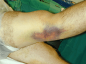
Inspection: Look for signs of external swelling and bruising or obvious deformity of the knee.
Ligament Laxity: Use the Lachman maneuver and stress the knee in 0-30˚ in varus and valgus positions to determine the degree of instability. Three out of four ligament disruption should raise suspicion for occult knee dislocation. By lifting the leg by the patient’s heels, the knee may fall into hyperextension also suggesting an unstable knee.
Vascular Examination: Vascular injury to the popliteal artery is the most dreaded complication Palpation of the tibialis posterior and dorsalis pedis pulses is crucial. Absent or reduced pulses merit immediate attention. If a pulse deficit is identified, management should not be delayed by imaging. Reduction should be performed (if not already reduced) and vascular surgery consultation should be obtained if lack of a pulse persists despite reduction.
The ankle-brachial index (ABI) is also important in determining management. Patients with an ABI > or equal to 0.9 can be safely observed with serial ABI examinations. The combination of normal ABIs and pulses has a 100% sensitivity for identifying vascular injury. Serial testing should be performed every 2 to 4 hours by adequately trained personnel for 48-72 hours in order to appropriately rule out popliteal artery injury. Conversely, those with an ABI of < 0.9 warrant further imaging with CT angiogram and possible consultation with vascular surgeon. Early consultation is vital because after 8 hours of ischemia, more than 80% of cases will require an amputation.
Neurological Examination: The two nerves that pass through the popliteal fossa, the tibial and common peroneal nerves, can often be affected in knee dislocations. Common peroneal nerve injury will lead to foot drop or difficulty with foot dorsiflexion, while tibial nerve injury will affect the ability to plantar flex the foot or the toes.
Imaging
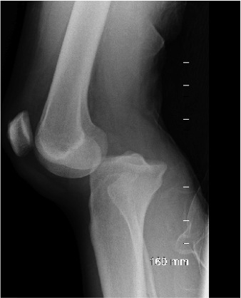
Plain radiographs should be obtained for all suspected dislocations. If pulses are absent or weak, reduction of the joint should not be delayed. Images of the knee may often be normal if the joint has reduced before the ED visit. Certain injuries such as a Segond fractures (avulsion fracture of the lateral tibial condyle from an ACL tear) or tibial plateau fracture can be seen with knee dislocations. In the dislocated joint, the radiograph can help identify the type of dislocation (anterior, posterior, lateral or medial) and guide management.
The gold standard for identifying popliteal injury is the CT angiogram. Though serial neurovascular exams are often sufficient in ruling out vascular compromise, CT angiography should be ordered in all patients with absent or reduced distal pulses post-reduction, ABI < 0.9 or any abnormality in the serial neurovascular exams. Though again, definitive management with limb saving surgery should not be delayed in order to obtain this test.
Management:
Reduction of the dislocated knee is usually easy given the laxity of the knee. Longitudinal traction of the tibia may be sufficient for the knee to reduce. If non-reducible, pressure can be applied in the opposite direction of the injury mechanism. For example, posterior dislocations are often from a direct blow to a flexed knee and the force required to reverse that mechanism would be to place pressure over the femur and then displace the tibia anteriorly. Conversely, in anterior dislocations, the tibia would have to be directed posteriorly. After the reduction, the affected limb should be splinted at 20-30˚ flexion.
Given the precarious nature of this injury, orthopedics consultants should be made aware of any suspected knee dislocations while in the ED for advice on management, serial examination and further follow-up. Open reduction may be necessary if multiple attempts at closed reduction fail and most will ultimately require operative repair of their multiple ligamentous injuries. In addition, vascular surgery should promptly be consulted if there is any sign of vascular injury on exam or CT angiogram.
References:
- Helman, A. and Helman, A. (2019). Occult Knee Injuries Pearls and Pitfalls | Emergency Medicine Cases | EM Cases. [online] Emergency Medicine Cases. Available at: https://emergencymedicinecases.com/occult-knee-injuries/ [Accessed 18 Nov. 2019].
- Orthobullets.com. (2019). Knee Dislocation – Trauma – Orthobullets. [online] Available at: https://www.orthobullets.com/trauma/1043/knee-dislocation [Accessed 18 Nov. 2019].
- Core EM. (2019). True Knee + Patellar Dislocations. [online] Available at: https://coreem.net/core/true-knee-patellar-dislocations/ [Accessed 18 Nov. 2019].
- Tintinalli JE, Ma O, Yealy DM, Meckler GD, Stapczynski J, Cline DM, Thomas SH. eds. Tintinalli’s Emergency Medicine: A Comprehensive Study Guide, 9e New York, NY: McGraw-Hill; . http://accessemergencymedicine.mhmedical.com.proxy3.library.mcgill.ca/content.aspx?bookid=2353§ionid=183421313. Accessed November 15, 2019.
- Menaker Jay. Upper and Lower Extremity Long Bone Injury. In: Mattu A and Swadron S, ed. CorePendium. Burbank, CA: CorePendium, LLC. https://www.emrap.org/corependium/chapter/recVLRJPnSBgTJo9a/Upper-and-Lower-Extremity-Long-Bone-Injury#h.x6eybgmtfobb. Updated November 17, 2019. Accessed November 15, 2019.
- McClary, Kaylan N. “Ankle, Brachial Index (ABI).” StatPearls [Internet]., U.S. National Library of Medicine, 26 June 2019, http://www.ncbi.nlm.nih.gov/books/NBK544226/#__NBK544226_ai__.
Written by : Vanessa Knight, CCFP-EM resident, McGill University
Reviewed by Chanel Fortier-Tougas
